PREVENTION & THERAPY
Decoding Your Diagnosis: A Patient’s Guide to Orthopedic X-rays, MRI, Ultrasound, and CT Scans

Orthopedic imaging tests can be confusing, especially when dealing with pain. You might wonder if you need an X-ray or an MRI, what the scan will show, or why your doctor picked a certain test.
This guide explains everything in simple terms.
We’ll walk you through the most common imaging options, what each one is used for, and what to expect, so you can feel more confident and informed about your care.
ATTENTION! If you’re considering a scan for your orthopedic condition, Dr. Badia and his team are here to provide expert insight. Schedule a consultation at the Badia Hand to Shoulder Center to gain clarity and confidence in your treatment path.
Why Imaging Matters in Orthopedic Diagnosis
Imaging helps your doctor see what’s really going on inside your body when you’re dealing with joint pain, stiffness, or injury.
While a physical exam is a good starting point, it doesn’t always give the full picture. That’s where imaging comes in. Whether it’s an X-ray, MRI, CT scan, or ultrasound, the right test can confirm a diagnosis, show the extent of an injury, or guide treatment.
Imaging takes out the guesswork and helps your care team make clear, informed decisions so you can get back to moving comfortably and confidently.
X-rays: Your First Step in Understanding Bone Injuries
X-rays are often the first imaging test used when you visit an orthopedic specialist. They are quick, safe, and provide a clear view of your bones and joints.
If you’ve had a fall, feel pain, or notice swelling, an X-ray can help reveal what’s going on inside.
What X-rays can help diagnose:
- Fractures and dislocations
- Signs of arthritis (joint space narrowing, bone spurs)
- Infections or bone lesions
- Bone tumors or abnormalities
Why doctors use X-rays:
- Fast results to guide next steps in care
- High detail for seeing bones clearly
- Simple, affordable, and widely available
What to expect:
- You may be asked to remove metal items
- The scan takes just a few minutes and doesn’t hurt
- You can go back to your normal routine right after
X-rays are a helpful starting point for understanding injuries and choosing the right treatment.
MRI: Detailed Views of Soft Tissues and Joints
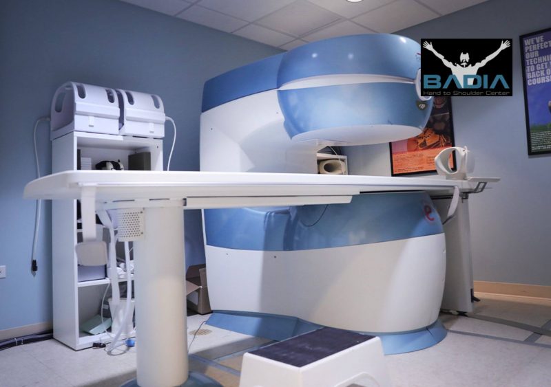
MRI (Magnetic Resonance Imaging) offers detailed cross-sectional images of your body using magnets and radio waves while radiation is not involved.
What MRIs are commonly used for:
- Rotator cuff or labral tears in the shoulder
- ACL or meniscus injuries in the knee
- Tendon and ligament damage
- Detecting arthritis, inflammation, tumors, or infections
What to expect:
- You will lie still on a table that slides into a tunnel-shaped scanner
- The scan takes about 30 to 60 minutes
- Some machines can feel enclosed, but open MRI options may be available
- The machine makes loud tapping sounds, so ear protection is usually provided
MRIs are safe for most people, though patients with certain metal implants or pacemakers may need special evaluation before the scan.
Ultrasound: Real-Time Imaging for Tendons and Muscles
Ultrasound uses high-frequency sound waves to produce live images of your body’s soft tissues. It’s a safe, comfortable option, especially helpful when your symptoms change with movement.
What ultrasound can help with:
- Diagnosing tendon tears or inflammation
- Spotting fluid buildup or bursitis
- Evaluating soft tissues during movement
- Guiding joint injections with greater accuracy
- Providing real-time feedback so the doctor can explain what they’re seeing during the scan
What to expect:
- A handheld device glides over your skin with gel
- No pain, radiation, or need for contrast dye
- Results may be available immediately
Ultrasound is especially helpful because it allows movement during the test. It is quick, non-invasive, and does not require tight spaces or contrast dye. Many patients find it comfortable, convenient, and helpful for understanding their condition on the spot.
CT Scans: Comprehensive 3D Views for Complex Cases
When a regular X-ray isn’t enough, a CT scan may be ordered. CT (computed tomography) combines multiple X-rays to create detailed 3D images of bones, joints, and soft tissues.
CT scans help diagnose:
- Complex fractures
- Subtle bone abnormalities
- Joint damage that needs surgical planning
- Internal bleeding or infections after injury
What to expect:
- You will lie on a table that moves slowly through a round scanner
- The test is painless and usually takes just a few minutes
- You may be asked to stay very still, and sometimes a contrast dye is used to highlight certain areas
CT scans involve more radiation than an X-ray, but they provide a far deeper view, especially when surgery is on the table.
How Your Doctor Chooses the Right Scan
- Take microbreaks every 30 to 45 minutes
Brief pauses help reset your posture, improve blood flow, and reduce stiffness. Stand up, stretch, or walk for a minute. Even small movements can make a big difference over time. - Stretch your wrists and forearms regularly
Long hours of typing can strain your forearms and wrists. To relieve tension, extend one arm with your palm facing up and gently pull your fingers back with the other hand. Hold for 30 seconds and switch sides. - Incorporate shoulder and neck mobility exercises
Sitting too long can lead to tight shoulders and a stiff neck. Shrug your shoulders up and down 10 times, then roll your neck slowly in each direction to release built-up tension. - Pair movement with daily routines
Stand during phone calls, do a few stretches while waiting for a file to load, or walk around after meetings. Linking movement with daily tasks helps you build healthy habits naturally.
When figuring out the best scan, your doctor considers your symptoms, medical history, and exam results. They’re not just picking randomly.
- X-rays are ideal for bones and joints
- MRI is best for ligaments, cartilage, and nerves
- CT gives in-depth views when more detail is needed
- Ultrasound captures soft tissues in motion
Other factors like whether you have implants, are pregnant, or are claustrophobic, also guide the decision.
Your provider’s goal is to choose the safest, most accurate test to get answers quickly.
Preparing for Your Imaging Appointment
Feeling nervous? That’s normal. A little preparation can help ease your mind and make the appointment go smoothly.
Before your visit:
- Arrive 15-30 minutes early for check-in
- Bring ID, insurance card, and past imaging records
- Ask in advance about fasting or medication instructions
- Tell your provider if you’re pregnant or have implants
What to wear:
- Comfortable, loose clothing
- Avoid metal (zippers, underwire bras, jewelry)
If contrast dye is needed, you may be asked about allergies or kidney function.
Always speak up if you’re unsure, your care team is there to help.
Understanding your diagnostic images is a crucial step in your health journey.
If you have questions about your orthopedic imaging results or need clarification on your diagnosis, don’t hesitate to reach out to Dr. Badia and the Badia Hand to Shoulder Center.
Your care team is there to guide you through the process.
If you’re ever unsure, don’t hesitate to ask questions. They want you to feel safe, informed, and supported every step of the way.
References
- Aagesen, A. L., & Melek, M. (2020). Choosing the right diagnostic imaging modality in musculoskeletal diagnosis.
- American College of Radiology. (n.d.). RadiologyInfo: Your radiologist explains medical imaging procedures.
- National Institute of Biomedical Imaging and Bioengineering. (2022). Ultrasound. U.S. Department of Health & Human Services.
- Radiological Society of North America. (2023). CT (Computed Tomography) scan. RadiologyInfo.org.


