
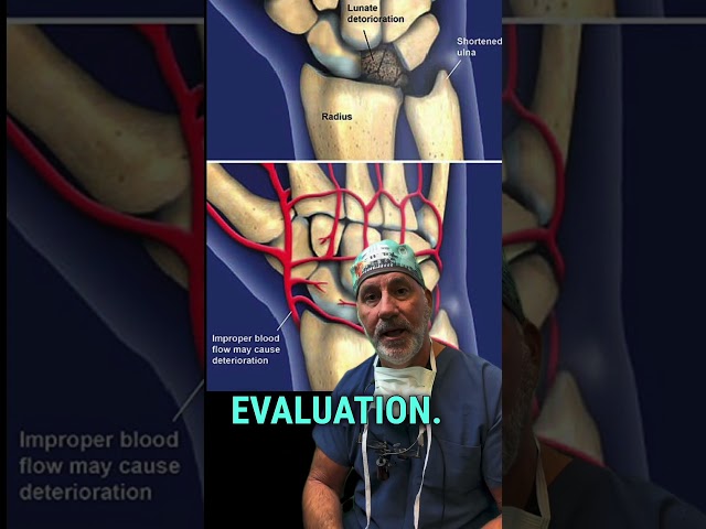


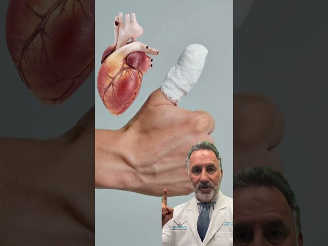
Patient from Ecuador with an injury to the capitohamate (proximal) ligament and the lunotriquetral ligament.
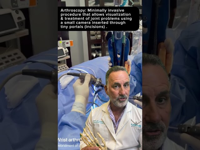
The arthroscopy images show a significant gap and instability between the capitate and hamate bones. These injuries are difficult to see with an MRI, and in this patient’s case, the MRI did not demonstrate the injury.
The first image shows passage through the lunotriquetral ligament interval, confirming a tear of the critical ligament on the ulnar or pinky side of the wrist.
This is a sign of an extensive injury to the ulnar column of the wrist that is often seen in sports injuries and industrial trauma.
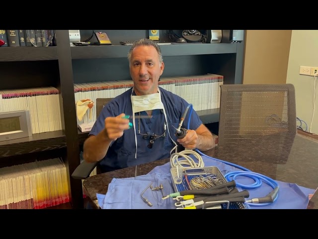
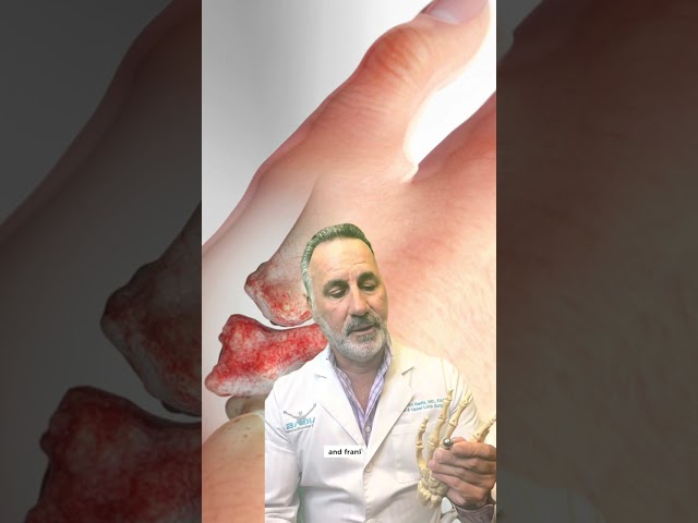
Arthroscopic debridement of the ligaments is performed to stimulate the healing of a new ligament, which is accelerated by the injection of an orthobiologic.
Stem cells or PRP and pins are placed under the skin and are removed approximately seven weeks after surgery, at which time therapy and gradual return to sports or work begins.